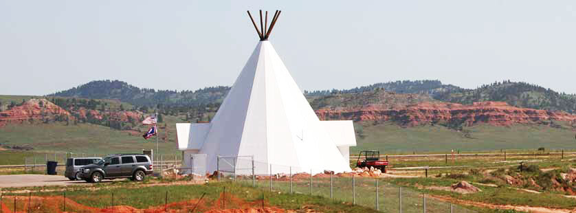
Labor Day, 5 September will be the last day we will be open this summer. Come out, visit us and celebrate another year!

Labor Day, 5 September will be the last day we will be open this summer. Come out, visit us and celebrate another year!

Contact Information
Vore Buffalo Jump Foundation P.O. Box 369 Sundance, WY 82729 Telephone: (307) 266-9530Contact Information
Vore Buffalo Jump Foundation
P.O. Box 369
Sundance, WY 82729
Telephone: (307) 266-9530
Our email address is: info@vorebuffalojump.org
Check out our Facebook Feed! Like our page so you don't miss current updates.
This week, Mariah Newtson, who is a UWAR curation technician, shares an artifact. The photographs shown are of a bison bone from the Vore Site that has been etched by plant roots. The photographs were taken with the DinoLite Digital Microscope. Root etching can mar the surfaces of human or faunal remains found in archaeological sites. Plant roots are acidic, and when they come into contact with bone, they begin to decalcify the surface. This can result in the appearance of an engraving or imprint of the roots on the surface of the bone. At the Vore site, these etchings would have been due to roots of shortgrass, yucca, poison ivy, stinging nettles, or chokecherry. Archaeologists care about root etching because it is a taphonomic process. These are processes that occur after an organism’s death, and they often modify remains in ways that can interfere with archaeological interpretation and research. The knowledge of whether or not a bone modification on an artifact was the result of human interaction influences the cultural relevance of the element. Thus, it is important to understand root etching so that it is not confused with human activities.
Research of the Vore collection is funded in part by a grant from the Institute of Museum and Library Services and in part by donations from supporters to the Vore Buffalo Jump Foundation.
... See MoreSee Less


- Likes: 17
- Shares: 2
- Comments: 0
This week, Vore Scholar Dakota Buhmann is sharing an interesting pathology in mandibles from the Vore Collection. Pictured are the teeth of mandibles from two different adult bison. The fully erupted 3rd molar means that these animals were at least 3.5 years of age. As bison age, constant chewing wears down their teeth. These two individuals show unusual wear patterns. Molars 1 and 2 exhibit severe signs of wear while the 3rd molar shows significantly less wear, as all tooth cusps are visible and distinct. Though more wear is expected on molars 1 and 2 since they erupt before 3, the reason for this wear discrepancy in different individuals is unknown. These bison might have been born and raised for the first couple years of their lives in an area with tougher, more silica-rich grasses before moving to a different area. Perhaps there was a severe drought during their first couple years of life that incorporated more grit into their diets. Let us know what you think!
Research of the Vore collection is funded in part by a grant from the Institute of Museum and Library Services and in part by donations from supporters to the Vore Buffalo Jump Foundation.
... See MoreSee Less


As good as they are about dating things it would be a real possibility through tree rings or some other method to cross that with carbon dating and hypothesize an exact year of the jump event.
Do they replace 1 & 2?
Vore Scholar Haley Purifoy presents this week’s artifact. Bones develop and grow as individuals age. First, specialized cells called chondrocytes deposit cartilage. As the individual ages, the cartilage is replaced with woven bone, which then solidifies into mature bone. In long bones, the growth of the bone occurs at the epiphyseal plate, the area between the diaphysis (which is the bone shaft) and the epiphysis (which is the bone end). After bone growth is complete, the epiphysis and the diaphysis fuse together into one solid bone. Pictured are some of the stages of growth for bison posterior (or hindleg) metapodials. Metapodials on a bison are equivalent to the metacarpals, the bones of the palm of the hand, and metatarsals, the bones of the middle of the foot, in humans. In bison, these are one bone. The distal, or bottom, end of the metapodials fuse last. In a bison, the metapodial epiphyses forms as two separate pieces that later combine into one. The distal epiphysis is originally two separate bone fragments that fuse together and then fuse to the diaphysis of the bone. Even in a fully fused bone, the line of epiphyseal union, commonly called the growth plate, can still be seen. The fusion process for metapodials begins at about the age of six and continues until about the age of nine for the bison.
Research of the Vore collection is funded in part by a grant from the Institute of Museum and Library Services and in part by donations from supporters to the Vore Buffalo Jump Foundation.
... See MoreSee Less


The weather disrupted our usual artifact of the week program, so I'm sharing some photos of one of the special exhibits at the Vore Site. We are honored to display a bison hide tipi made by students from Chief Dull Knife College in 2014. The six hides that make up the tipi were tanned using bison brain and lots of hard work and were stitched together with sinew. ... See MoreSee Less


This week Molly Herron is sharing a canid-canine comparison – try saying that five times in a row! Canids (coyotes, wolves, dogs, etc.) have four canine teeth in their dentition. These teeth are vital for catching prey and holding on tightly as they sink into the flesh. Like all aspects of anatomy, form follows function – therefore, the maxillary (upper) and mandibular (lower) canine teeth are shaped differently. The maxillary canines are thicker and shorter, and the mandibular canines are narrower and longer. The mandibular canines also have a lingual ridge that runs along the inside of the tooth (indicated by the red arrow); this ridge helps to slice into flesh and gives the canid a better grip. Additionally, the maxillary and mandibular canines must have different shapes to fit together in the dog’s mouth – the space where the canines overlap is called the diastema. Although these two canine teeth from the Vore Bison Sinkhole site are not from the same animal, they clearly demonstrate this difference.
Research of the Vore collection is funded in part by a grant from the Institute of Museum and Library Services and in part by donations from supporters to the Vore Buffalo Jump Foundation.
... See MoreSee Less


Send us a message. Fill out the form and click on the submit button.
Check out our Facebook Feed! Like our page so you don't miss current updates.
This week, Mariah Newtson, who is a UWAR curation technician, shares an artifact. The photographs shown are of a bison bone from the Vore Site that has been etched by plant roots. The photographs were taken with the DinoLite Digital Microscope. Root etching can mar the surfaces of human or faunal remains found in archaeological sites. Plant roots are acidic, and when they come into contact with bone, they begin to decalcify the surface. This can result in the appearance of an engraving or imprint of the roots on the surface of the bone. At the Vore site, these etchings would have been due to roots of shortgrass, yucca, poison ivy, stinging nettles, or chokecherry. Archaeologists care about root etching because it is a taphonomic process. These are processes that occur after an organism’s death, and they often modify remains in ways that can interfere with archaeological interpretation and research. The knowledge of whether or not a bone modification on an artifact was the result of human interaction influences the cultural relevance of the element. Thus, it is important to understand root etching so that it is not confused with human activities.
Research of the Vore collection is funded in part by a grant from the Institute of Museum and Library Services and in part by donations from supporters to the Vore Buffalo Jump Foundation.
... See MoreSee Less


This week, Vore Scholar Dakota Buhmann is sharing an interesting pathology in mandibles from the Vore Collection. Pictured are the teeth of mandibles from two different adult bison. The fully erupted 3rd molar means that these animals were at least 3.5 years of age. As bison age, constant chewing wears down their teeth. These two individuals show unusual wear patterns. Molars 1 and 2 exhibit severe signs of wear while the 3rd molar shows significantly less wear, as all tooth cusps are visible and distinct. Though more wear is expected on molars 1 and 2 since they erupt before 3, the reason for this wear discrepancy in different individuals is unknown. These bison might have been born and raised for the first couple years of their lives in an area with tougher, more silica-rich grasses before moving to a different area. Perhaps there was a severe drought during their first couple years of life that incorporated more grit into their diets. Let us know what you think!
Research of the Vore collection is funded in part by a grant from the Institute of Museum and Library Services and in part by donations from supporters to the Vore Buffalo Jump Foundation.
... See MoreSee Less


As good as they are about dating things it would be a real possibility through tree rings or some other method to cross that with carbon dating and hypothesize an exact year of the jump event.
Do they replace 1 & 2?
Vore Scholar Haley Purifoy presents this week’s artifact. Bones develop and grow as individuals age. First, specialized cells called chondrocytes deposit cartilage. As the individual ages, the cartilage is replaced with woven bone, which then solidifies into mature bone. In long bones, the growth of the bone occurs at the epiphyseal plate, the area between the diaphysis (which is the bone shaft) and the epiphysis (which is the bone end). After bone growth is complete, the epiphysis and the diaphysis fuse together into one solid bone. Pictured are some of the stages of growth for bison posterior (or hindleg) metapodials. Metapodials on a bison are equivalent to the metacarpals, the bones of the palm of the hand, and metatarsals, the bones of the middle of the foot, in humans. In bison, these are one bone. The distal, or bottom, end of the metapodials fuse last. In a bison, the metapodial epiphyses forms as two separate pieces that later combine into one. The distal epiphysis is originally two separate bone fragments that fuse together and then fuse to the diaphysis of the bone. Even in a fully fused bone, the line of epiphyseal union, commonly called the growth plate, can still be seen. The fusion process for metapodials begins at about the age of six and continues until about the age of nine for the bison.
Research of the Vore collection is funded in part by a grant from the Institute of Museum and Library Services and in part by donations from supporters to the Vore Buffalo Jump Foundation.
... See MoreSee Less


The weather disrupted our usual artifact of the week program, so I'm sharing some photos of one of the special exhibits at the Vore Site. We are honored to display a bison hide tipi made by students from Chief Dull Knife College in 2014. The six hides that make up the tipi were tanned using bison brain and lots of hard work and were stitched together with sinew. ... See MoreSee Less


This week Molly Herron is sharing a canid-canine comparison – try saying that five times in a row! Canids (coyotes, wolves, dogs, etc.) have four canine teeth in their dentition. These teeth are vital for catching prey and holding on tightly as they sink into the flesh. Like all aspects of anatomy, form follows function – therefore, the maxillary (upper) and mandibular (lower) canine teeth are shaped differently. The maxillary canines are thicker and shorter, and the mandibular canines are narrower and longer. The mandibular canines also have a lingual ridge that runs along the inside of the tooth (indicated by the red arrow); this ridge helps to slice into flesh and gives the canid a better grip. Additionally, the maxillary and mandibular canines must have different shapes to fit together in the dog’s mouth – the space where the canines overlap is called the diastema. Although these two canine teeth from the Vore Bison Sinkhole site are not from the same animal, they clearly demonstrate this difference.
Research of the Vore collection is funded in part by a grant from the Institute of Museum and Library Services and in part by donations from supporters to the Vore Buffalo Jump Foundation.
... See MoreSee Less